







Look inside the brain
The brain is like a group of experts. All the parts of the brain work together, but each part has its own special responsibilities.
Source: National Institute of Neurological Disorders and Stroke, Brain Basics: Know Your Brain.
Clinic
3934 Union Rd, St. Louis, MO 63125.
Mon-Thu 7:00am-6:00pm, Fri 7:00am-2:00pm, closed Sat and Sun.
Featured articles

Scroll to look inside
Skip the tourIllustrative anatomy. Not a patient scan and not a diagram of how any treatment works. Credits
Anatomy: Z-Anatomy / BodyParts3D (DBCLS), adapted via Brain Project · CC BY-SA 4.0 · Credits
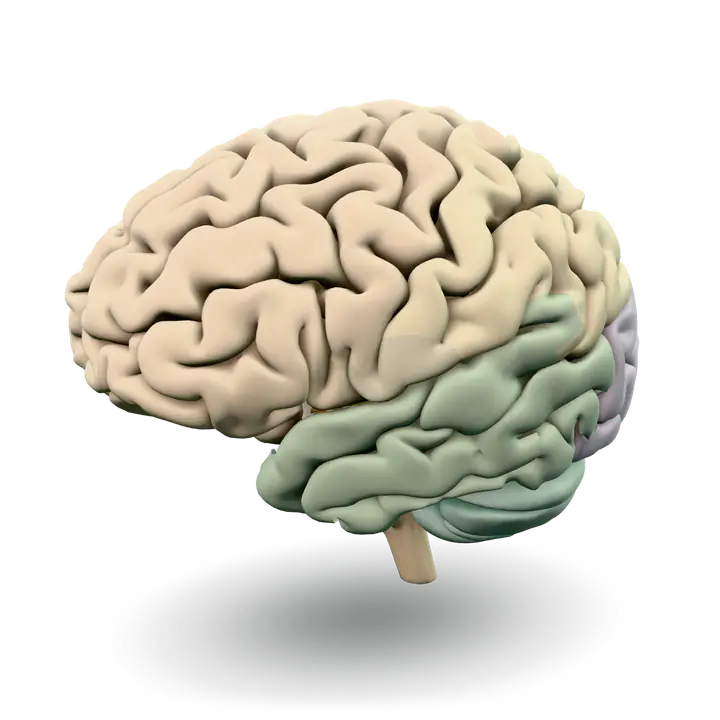
The brain is like a group of experts. All the parts of the brain work together, but each part has its own special responsibilities.
Source: National Institute of Neurological Disorders and Stroke, Brain Basics: Know Your Brain.
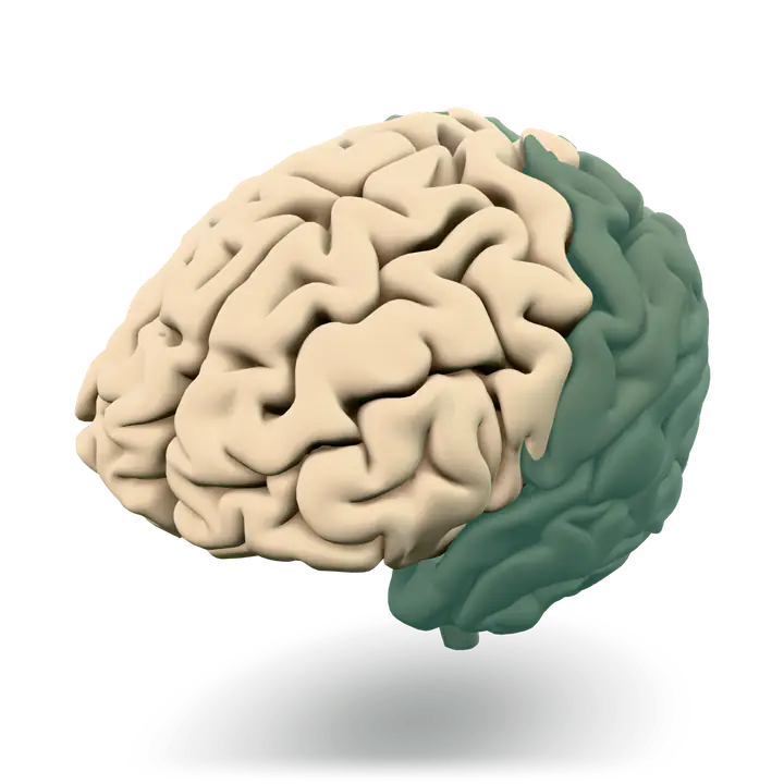
The two frontal lobes lie directly behind the forehead. When you plan a schedule, imagine the future, or use reasoned arguments, these two lobes do much of the work.
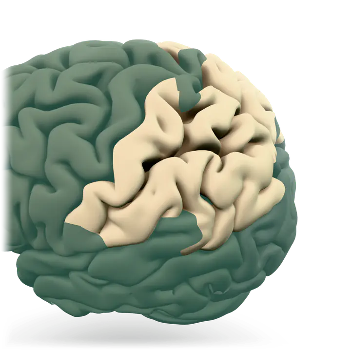
When you enjoy a good meal, the taste, smell, and texture of the food, two sections behind the frontal lobes called the parietal lobes are at work. The parietal lobes also support reading and arithmetic.
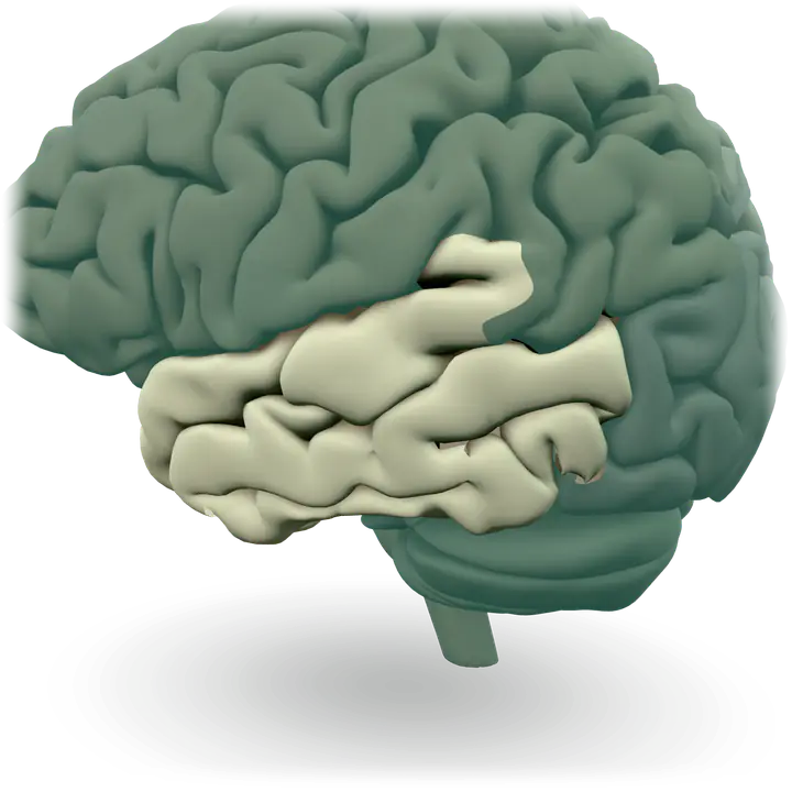
At the top of each temporal lobe is an area responsible for receiving information from the ears. The underside of each temporal lobe plays a crucial role in forming and retrieving memories, including those associated with music.
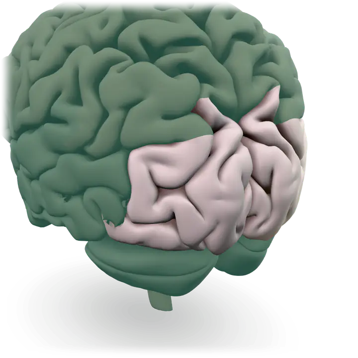
As you look at the words and pictures on this page, two areas at the back of the brain are at work. These lobes, called the occipital lobes, process images from the eyes and link that information with images stored in memory.
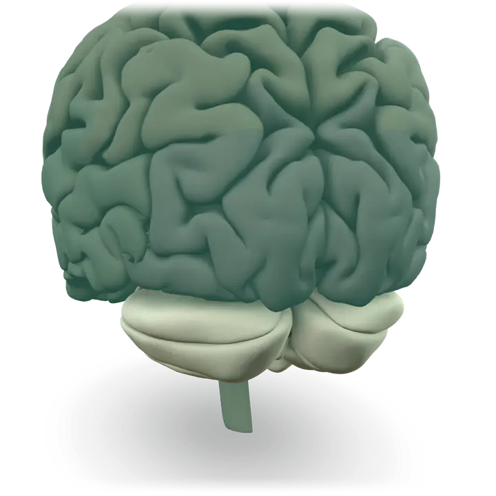
The cerebellum coordinates movement and is involved in learned movements. When you play the piano or hit a tennis ball, you are activating the cerebellum.
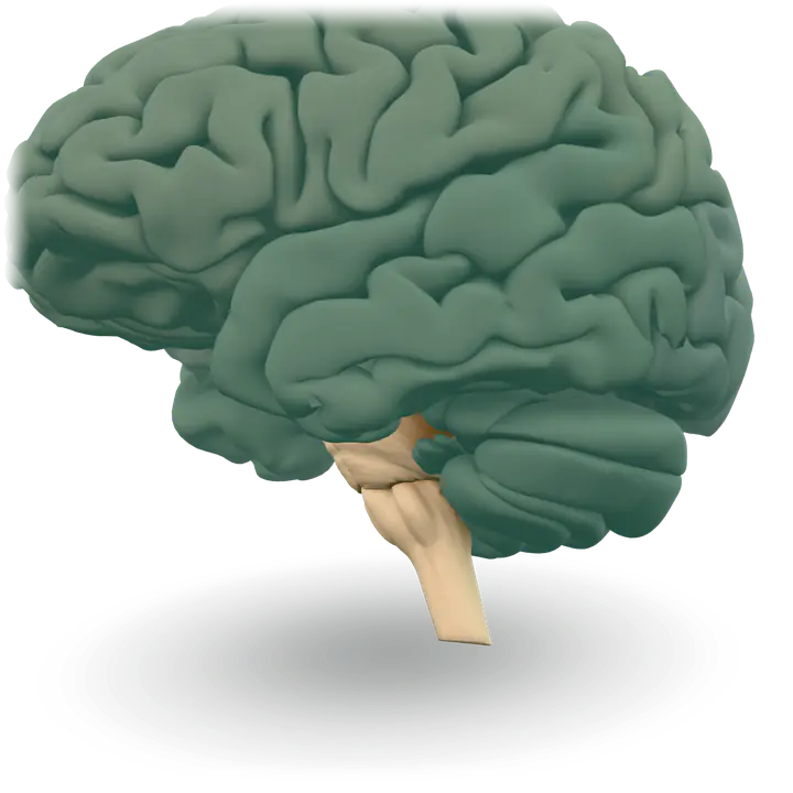
The hindbrain includes the upper part of the spinal cord, the brain stem, and a wrinkled ball of tissue called the cerebellum. The hindbrain controls the body’s vital functions such as respiration and heart rate.
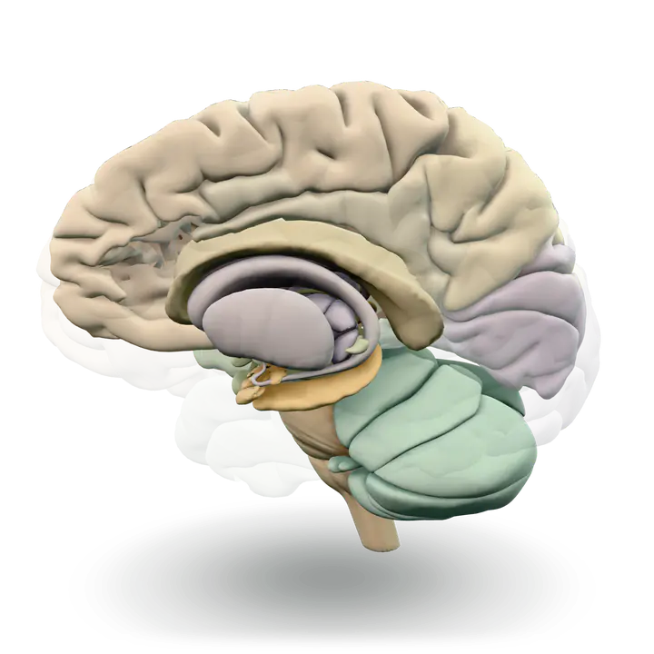
Deep within the brain, hidden from view, lie structures that are the gatekeepers between the spinal cord and the cerebral hemispheres.
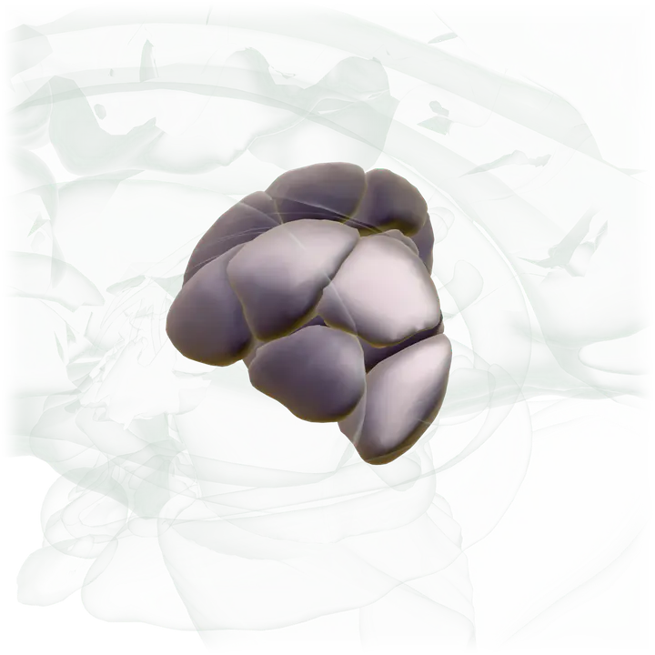
Near the hypothalamus lies the thalamus, a major clearinghouse for information going to and from the spinal cord and the cerebrum.
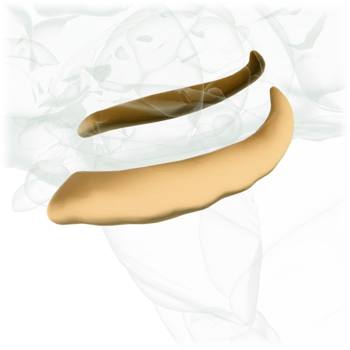
An arching tract of nerve cells leads from the hypothalamus and the thalamus to the hippocampus. This tiny nub acts as a memory indexer, sending memories out to the appropriate part of the cerebral hemisphere for long-term storage and retrieving them when necessary.
Start with the frontal lobe, or drag to turn the brain.
Illustrative anatomy. Not a patient scan and not a diagram of how any treatment works.
Anatomy: Z-Anatomy / BodyParts3D (DBCLS), adapted via Brain Project · CC BY-SA 4.0 · Credits
The brain is like a group of experts. All the parts of the brain work together, but each part has its own special responsibilities.
The two frontal lobes lie directly behind the forehead. When you plan a schedule, imagine the future, or use reasoned arguments, these two lobes do much of the work.
When you enjoy a good meal, the taste, smell, and texture of the food, two sections behind the frontal lobes called the parietal lobes are at work. The parietal lobes also support reading and arithmetic.
At the top of each temporal lobe is an area responsible for receiving information from the ears. The underside of each temporal lobe plays a crucial role in forming and retrieving memories, including those associated with music.
As you look at the words and pictures on this page, two areas at the back of the brain are at work. These lobes, called the occipital lobes, process images from the eyes and link that information with images stored in memory.
The cerebellum coordinates movement and is involved in learned movements. When you play the piano or hit a tennis ball, you are activating the cerebellum.
The hindbrain includes the upper part of the spinal cord, the brain stem, and a wrinkled ball of tissue called the cerebellum. The hindbrain controls the body’s vital functions such as respiration and heart rate.
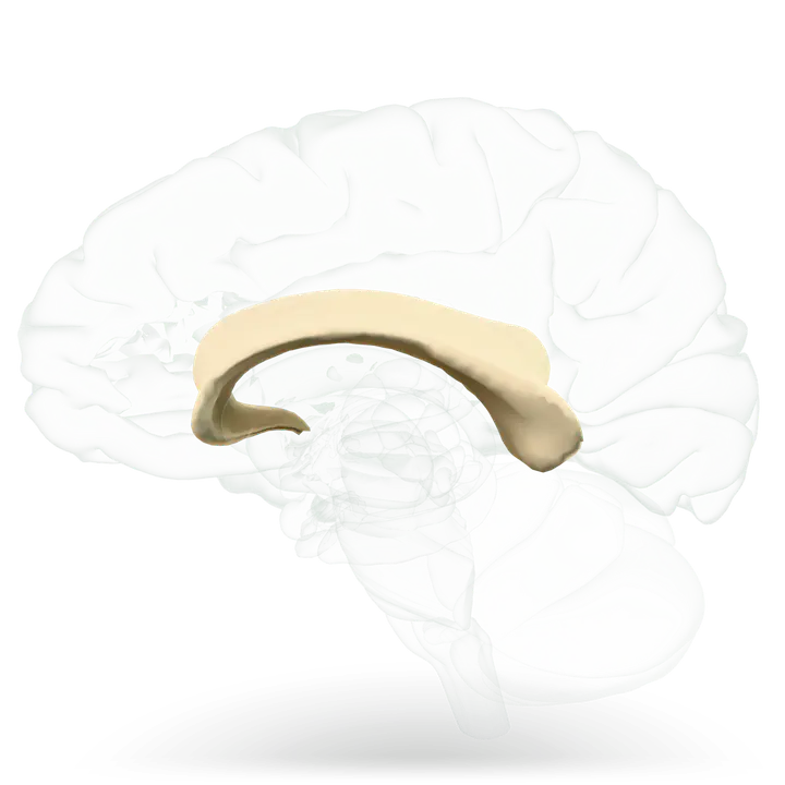
The cerebrum is split into two halves (hemispheres) by a deep fissure. The two cerebral hemispheres communicate with each other through a thick tract of nerve fibers that lies at the base of this fissure, called the corpus callosum.
Near the hypothalamus lies the thalamus, a major clearinghouse for information going to and from the spinal cord and the cerebrum.
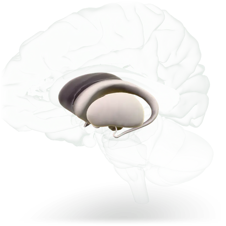
The basal ganglia are clusters of nerve cells surrounding the thalamus. They are responsible for initiating and integrating movements.
An arching tract of nerve cells leads from the hypothalamus and the thalamus to the hippocampus. This tiny nub acts as a memory indexer, sending memories out to the appropriate part of the cerebral hemisphere for long-term storage and retrieving them when necessary.
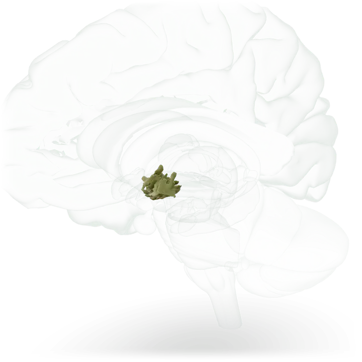
The hypothalamus, about the size of a pearl, directs a multitude of important functions. It wakes you up in the morning and gets the adrenaline flowing during a test or job interview.
Source: National Institute of Neurological Disorders and Stroke, Brain Basics: Know Your Brain.
The brain model is from Z-Anatomy, built on BodyParts3D by the Database Center for Life Science (DBCLS), as shared by the Brain Project. It is published under the Creative Commons Attribution-ShareAlike 4.0 license. We adapted it for this website, and the adapted model and its pictures carry the same license.